Research Overview · Q2-2026
Dehydroepiandrosterone (DHEA): Chemical Identity & Anticancer Potential
A comprehensive examination of DHEA's molecular notation, structural features, and emerging roles as an anticancer agent across breast, colorectal, and bladder cancers — with mechanistic comparison.
Reference — Wikipedia
Dehydroepiandrosterone (DHEA)
Dehydroepiandrosterone (DHEA), also known as androstenolone, is an endogenous steroid hormone precursor and one of the most abundant circulating steroids in humans. It is produced in the adrenal glands, gonads, and brain, functions as a metabolic intermediate in sex steroid biosynthesis, and has direct biological effects via nuclear receptors, neurosteroid activity, and metabolic enzyme modulation.
Pharmacology
Bioavailability (oral)
~50%
Half-life (DHEA)
~25 min
Half-life (DHEA-S)
~11 h
Routes
Oral, vaginal, IM
Biological Functions
As an androgen
Adrenal androgens including DHEA drive adrenarche (pubic/axillary hair, body odor, skin oiliness). DHEA is potentiated locally to testosterone and DHT in skin and hair follicles.
As an estrogen
DHEA is a weak estrogen and is converted to potent estrogens such as estradiol in tissues like the vagina, producing estrogenic effects locally.
As a neurosteroid
In the central nervous system, DHEA acts as a neurosteroid and neurotrophin modulator with important effects on neuronal survival and signaling.
Key Biological Activities
- Androgen receptor: Weak partial agonist (Ki ≈ 1 μM); activity is context-dependent relative to circulating testosterone/DHT.
- Estrogen receptors: Binds ERα and ERβ; can act as a full agonist of ERβ at physiological concentrations.
- G6PD inhibition: Uncompetitive inhibitor of glucose-6-phosphate dehydrogenase (Ki ≈ 17 μM), lowering NADPH — relevant to oxidative stress and anticancer mechanisms.
- Neurotrophin receptors: DHEA and DHEA-S bind TrkA, p75NTR, TrkB, and TrkC with nanomolar affinity, acting as endogenous "steroidal microneurotrophins."
DHEA and Aging
DHEA peaks in early adulthood and declines with age. Supplementation is studied for restoring hormone levels and potential effects on energy, mood, libido, and bone density via androgenic metabolites and osteoblast/IGF-1 pathways.
Reference Serum Levels
Circulating DHEA declines from a peak around age 20. DHEA-S levels are typically 250–300× higher than DHEA and serve as a circulating reservoir.
| Population | DHEA (ng/dL) |
|---|---|
| Adult men | 180–1250 |
| Adult women | 130–980 |
| Pregnant women | 135–810 |
| Children (6–12 y) | 11–186 |
| Adolescent boys (Tanner IV–V) | 100–400 |
| Adolescent girls (Tanner IV–V) | 165–690 |
Biosynthesis & Distribution
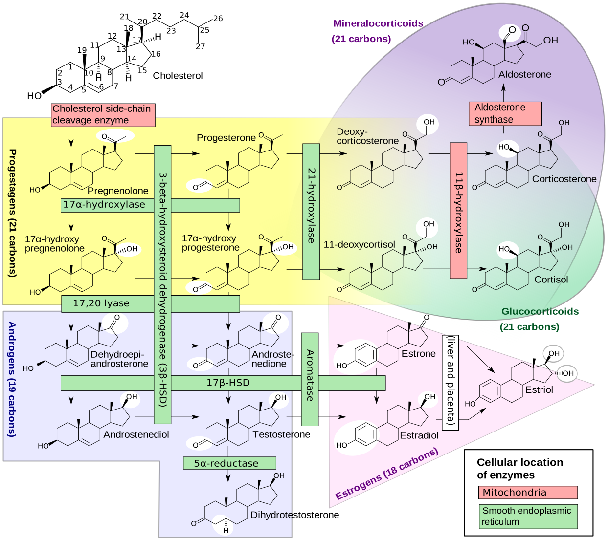
DHEA is synthesized in the zona reticularis of the adrenal cortex (ACTH-regulated) and in gonads (GnRH-regulated), from cholesterol via pregnenolone and 17α-hydroxypregnenolone (CYP11A1, CYP17A1). ~50–70% of circulating DHEA originates from peripheral desulfation of DHEA-S.
Metabolism
DHEA is sulfated to DHEA-S by SULT2A1/SULT1E1; DHEA-S levels are ~250–300× DHEA and can be converted back via steroid sulfatase. Terminal half-life: DHEA 15–30 min; DHEA-S 7–10 h. Metabolites include androstenediol, androstenedione, 7-keto-DHEA, and hydroxylated derivatives.
History
DHEA was first isolated from human urine in 1934 by Adolf Butenandt and Kurt Tscherning. In the United States it is sold as an over-the-counter supplement and as the prescription drug prasterone.
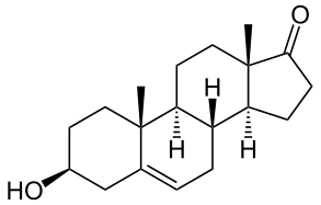
Also known as androst-5-en-3β-ol-17-one (δ5-epiandrosterone), a naturally occurring androstane 17-ketosteroid closely related to androstenediol, androstenedione, and testosterone.
Read full article on WikipediaSummarized from Wikipedia (CC BY-SA 4.0). Images from Wikimedia Commons under their respective licenses.
01 — Chemical Identity
Molecular Notation: SMILES & InChI
Every small molecule in drug discovery and cheminformatics is assigned two standard machine-readable identifiers: a SMILES string for structural encoding and an InChI identifier for uniqueness and cross-database linking. Understanding both is essential for in-silico docking, database queries, and computational pharmacology.
SMILES
[C@@H]1(CC[C@H]2[C@@H]1CC[C@H]3[C@@H]2CC(=O)[C@@]3(C)CC)OCanonical isomeric SMILES encoding absolute stereochemistry at six chiral centers.
InChI
InChI=1S/C19H28O2/c1-18-9-7-13(20)11-12(18)3-4-14-15-5-6-17(21)19(15,2)10-8-16(14)18/h3,13-16,20H,4-11H2,1-2H3/t13-,14+,15+,16+,18+,19+/m1/s1Standard InChI with stereochemistry layer. InChIKey: FMGSKLZLMKYGDP-USOAJAOKSA-N
What is SMILES?
SMILES (Simplified Molecular Input Line Entry System) is a compact line notation for describing the structure of a molecule using short ASCII strings. Developed by Daylight Chemical Information Systems, SMILES encodes atoms, bonds, branches, rings, and stereochemistry in a human-readable yet machine-parseable format.
SMILES Key Conventions
[C@@H]1(…)O
Tetrahedral chiral center (3β-OH group) — the defining hydroxyl of DHEA.
CC(=O)
17-ketone group at D-ring. Distinguishes DHEA from androstenediol.
C1CCC2…
Four fused carbocyclic rings (A/B/C/D) of the steroidal androstane skeleton.
@@, @ symbols
Six absolute stereocenters defining the biologically active trans-fused conformation.
What is InChI?
InChI (IUPAC International Chemical Identifier) is a non-proprietary standard identifier developed by IUPAC and NIST. Unlike SMILES, InChI is canonical — there is exactly one InChI per unique molecule, regardless of how it is drawn or input.
InChI Layer Breakdown for DHEA
The InChIKey (FMGSKLZLMKYGDP-USOAJAOKSA-N) is a 27-character hash of the full InChI, suitable for web searches and database indexing.
- IUPAC Name(3S,8R,9S,10R,13S,14S)-3-hydroxy-10,13-dimethyl-…-17-one
- Molecular FormulaC₁₉H₂₈O₂
- Exact Mass288.2089 Da
- Molecular Weight288.43 g/mol
- Chiral Centers6 (all defined)
- Ring SystemAndrostane (A–B–C–D)
- Key Functional Groups3β-OH, Δ5, 17-ketone
- LogP (est.)3.06
- H-bond donors / acceptors1 / 2
- Rotatable bonds0
Interpreting the Structure
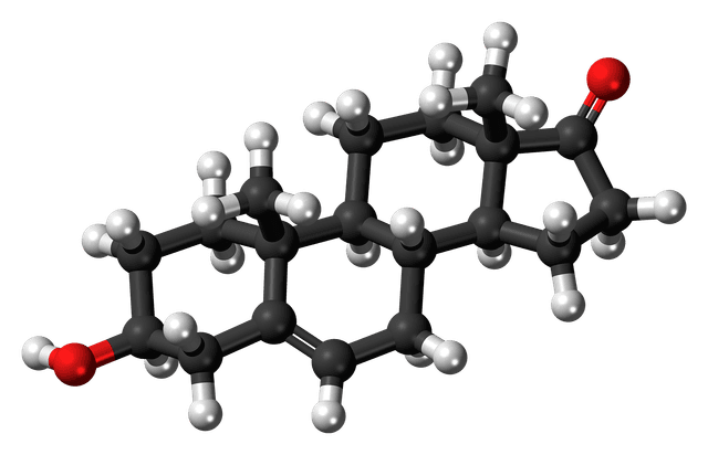
The skeletal diagram shows DHEA's four trans-fused rings (A–D). Ring A contains the 3β-hydroxyl group essential for biological activity and sulfation to DHEA-S. The A/B junction carries a Δ5 double bond. Ring D bears the 17-ketone, distinguishing DHEA from androgens like testosterone. Angular methyl groups (C18, C19) contribute to the rigid three-dimensional shape.
Research Assets & Action Center
1. Structural Data Downloads
2. Computational Chemistry Actions
02 — Biological Context
DHEA as a Biological Molecule
DHEA is the most abundant circulating steroid hormone in humans. It is produced primarily in the zona reticularis of the adrenal cortex, with secondary production in the gonads and brain. Serum concentration peaks in the mid-twenties and declines with age — adrenopause — making it a subject of intense interest in aging, oncology, and metabolic disease research.
DHEA itself is hormonally weak but serves as the primary circulating precursor for androgens and estrogens through peripheral conversion. Beyond hormone precursor roles, accumulating evidence shows direct effects: nuclear receptor binding, neurotrophin modulation, and inhibition of metabolic enzymes central to tumor survival.
Biosynthetic pathway: Cholesterol → Pregnenolone → DHEA (central node) → androgens and estrogens. Dashed arrow shows reversible sulfation to DHEA-S, the major circulating storage form.
03 — Oncological Potential
DHEA & Cancer: Mechanistic Evidence
Research spanning four decades has established dose-dependent antiproliferative, proapoptotic, and antimetastatic effects across multiple cancer types. The primary mechanism involves uncompetitive inhibition of glucose-6-phosphate dehydrogenase (G6PD) in the pentose phosphate pathway, alongside receptor-mediated signaling. The anticancer profile is cancer-type specific.
Core Anticancer Mechanisms of DHEA
DHEA exerts anticancer effects through several converging pathways. The most well-characterized is uncompetitive inhibition of G6PD, depleting cellular NADPH required for tumor biosynthesis, ROS detoxification, and carcinogen activation.
- G6PD Inhibition → Oxidative Stress: Reversible inhibition of mammalian G6PD lowers NADPH, reducing antioxidant capacity. Elevated ROS induces DNA and lipid damage, triggering apoptosis.
- Cell Cycle Arrest: DHEA induces G1-phase arrest, upregulating p21/p27 and reducing cyclin D1 expression.
- Apoptosis Induction: Caspase-3/9 activation, Bax/Bcl-2 modulation, and DR5 (TRAIL receptor) upregulation documented.
- Anti-inflammatory Action: Suppresses NF-κB signaling, reducing cytokines that sustain tumor microenvironments.
- Anti-proliferative & Anti-metastatic: Downregulates MMP-2/9 and suppresses WNT/β-catenin signaling in cancer stem cells.
- ERβ Agonism: Selective ERβ agonism activates tumor-suppressive signaling rather than proliferative ERα pathways.
04 — Comparative Analysis
Mechanistic Comparison Across Cancer Types
Current evidence on DHEA's anticancer activity across breast, colorectal, and bladder cancers — comparing mechanisms, evidence quality, p53 dependence, and research status.
| Parameter | Breast Cancer | Colorectal Cancer | Bladder Cancer |
|---|---|---|---|
| Primary Mechanism | G6PD inhibition; p16-senescence; p53 upregulation; ERβ agonism | ER stress → PERK/ATF4/CHOP → autophagy → apoptosis | G6PD inhibition → ROS accumulation → AKT suppression |
| p53 Dependence | Partial | Independent | Unknown |
| ER Status Sensitivity | Sensitive | Not applicable | Not applicable |
| In Vivo Evidence | Strong | Confirmed | Indirect |
| Effective In Vitro Dose | >10 μM (G6PD inhibition) | 50–400 μM across cell lines | Not directly quantified for DHEA |
| Key Signaling Nodes | G6PD · p53 · p16 · ERβ · Bcl-2 family | PERK · eIF2α · ATF4 · CHOP · ATG5 · DR5 · p21 | G6PD · NADPH · ROS · AKT · NF-κB |
| Combination Potential | Moderate | High | High |
| Clinical Translation | Preclinical | Early | Exploratory |
| Evidence Strength | Moderate–Strong | Strong | Moderate |
Key Distinctions
The most clinically significant distinction is DHEA's p53-independent mechanism in CRC. Because advanced CRC frequently carries p53 mutations, a drug killing cells regardless of p53 status has outsized translational value.
In breast cancer, antiproliferative effects occur at pharmacological concentrations exceeding normal tissue levels. DHEA can convert to estrogens, potentially stimulating ER-positive tumor growth — the most context-sensitive application.
For bladder cancer, mechanistic rationale is strong but direct DHEA evidence is thinner. G6PD overexpression combined with knockdown proof-of-concept makes this a high-priority research area.
Across all three cancer types, DHEA's multi-target profile contrasts with single-target chemotherapeutics — pleiotropy may reduce resistance emergence but complicates dose optimization.
05 — Research Framework
DARTS Protocol & In-Silico Context
The DHEA Research Portal's primary experimental framework — Drug Affinity Responsive Target Stability (DARTS) — is ideally suited to DHEA's multi-target profile. DARTS exploits drug-bound protein protection from proteolytic digestion; comparing band intensities after limited pronase digestion identifies novel binding partners without prior target knowledge.
The current protocol optimizes Pronase digestion ratios for HepG2 and HEK293T lysates, paired with LC-MS/MS on DARTS-stable bands. Integration with AutoDock Vina and SwissTargetPrediction (89.4% target prediction precision) creates a closed-loop discovery pipeline.
For cancer applications, DARTS is valuable because established targets (G6PD, PERK, estrogen receptors, androgen receptors) may represent only a subset of the binding interactome. Novel kinases and metabolic enzymes upregulated in CRC, breast, and bladder cells may emerge from unbiased profiling.